- Главная
- Разное
- Бизнес и предпринимательство
- Образование
- Развлечения
- Государство
- Спорт
- Графика
- Культурология
- Еда и кулинария
- Лингвистика
- Религиоведение
- Черчение
- Физкультура
- ИЗО
- Психология
- Социология
- Английский язык
- Астрономия
- Алгебра
- Биология
- География
- Геометрия
- Детские презентации
- Информатика
- История
- Литература
- Маркетинг
- Математика
- Медицина
- Менеджмент
- Музыка
- МХК
- Немецкий язык
- ОБЖ
- Обществознание
- Окружающий мир
- Педагогика
- Русский язык
- Технология
- Физика
- Философия
- Химия
- Шаблоны, картинки для презентаций
- Экология
- Экономика
- Юриспруденция
Что такое findslide.org?
FindSlide.org - это сайт презентаций, докладов, шаблонов в формате PowerPoint.
Обратная связь
Email: Нажмите что бы посмотреть
Презентация на тему Immunophysilogy of lung
Содержание
- 6. For more than 70 years, surfactant was
- 7. Augmented production of SP-A by the maturing
- 8. Surfactant protein A (SP-A) has been shown
- 9. A consequence of apoptotic-body uptake by a
- 10. The roles of macrophages in clearing apoptotic
- 11. The alveolar membrane is the largest surface
- 12. Alveolar macrophages are long-lived, with a turnover
- 13. The non-inflammatory clearance of apoptotic cells (a)
- 18. In healthy individuals, the airspaces are replete
- 19. Table 1 | The specific phenotype of
- 20. Innate immune functions of alveolar macrophages. As
- 21. Alveolar macrophages reside in the airspaces juxtaposed
- 22. Скачать презентацию
- 23. Похожие презентации
For more than 70 years, surfactant was perceived to be a soap-like substance that reduced surface tension in the lung and made breathing easier. With the advent of molecular techniques, it was discovered that one of

Слайд 7 Augmented production of SP-A by the maturing fetal
lung at term provides a key hormonal stimulus for
the cascade of inflammatory signaling pathways within the maternal uterus that culminate in the enhanced myometrial contractility leading to parturition. This hormonal signal, transmitted to the uterus by fetal AF macrophages reveals that the fetal lungs are sufficiently developed to withstand the critical transition from an aqueous to an aerobic environment.Increased amounts of surfactant lipids and proteins are secreted by the fetal lung into the amniotic fluid during the final third of gestation. Recent studies show that surfactant protein A (SP-A) acts on amniotic-fluid macrophages to induce their migration to the pregnant uterus and the
secretion of inflammatory mediators. This results to initiation of parturition.
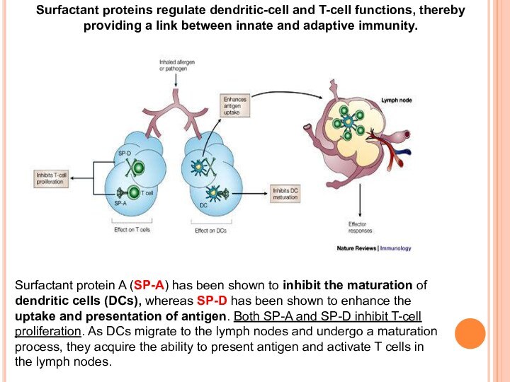
Слайд 8 Surfactant protein A (SP-A) has been shown to
inhibit the maturation of dendritic cells (DCs), whereas SP-D
has been shown to enhance the uptake and presentation of antigen. Both SP-A and SP-D inhibit T-cell proliferation. As DCs migrate to the lymph nodes and undergo a maturation process, they acquire the ability to present antigen and activate T cells in the lymph nodes.Surfactant proteins regulate dendritic-cell and T-cell functions, thereby providing a link between innate and adaptive immunity.
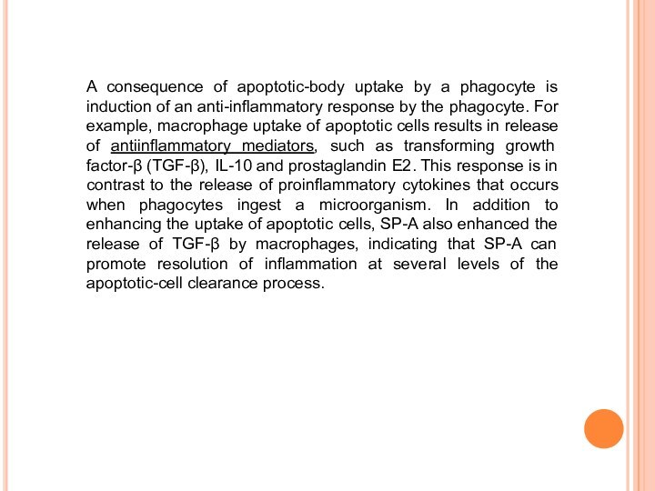
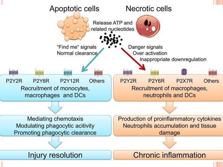
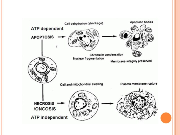
Слайд 9 A consequence of apoptotic-body uptake by a phagocyte
is induction of an anti-inflammatory response by the phagocyte.
For example, macrophage uptake of apoptotic cells results in release of antiinflammatory mediators, such as transforming growth factor-β (TGF-β), IL-10 and prostaglandin E2. This response is in contrast to the release of proinflammatory cytokines that occurs when phagocytes ingest a microorganism. In addition to enhancing the uptake of apoptotic cells, SP-A also enhanced the release of TGF-β by macrophages, indicating that SP-A can promote resolution of inflammation at several levels of the apoptotic-cell clearance process.Слайд 10 The roles of macrophages in clearing apoptotic cells
and cellular debris in health and disease are equally
as important as the participation of these cells in immunological responses. However, each function requires plasticity within the resident macrophage population so that pro-inflammatory responses to tissue debris or to innocuous antigens are inhibited, but effective immune responses to pathogenic microorganisms are not compromised. The ability of tissue macrophages to adapt and to carry out such disparate functions led to their broad classification as either classically activated M1 macrophages or alternatively activated M2 macrophages. Since their initial description, the functional and phenotypical characteristics of macrophages within the M1 phenotype have remained mostly unaltered, but the M2 macrophage category has been expanded to accommodate a broad range of macrophage functions in wound healing and in immune regulation. A transcriptional analysis of human alveolar macrophages that were polarized ex vivo using interferon‑γ (IFNγ), or with interleukin‑4 (IL‑4) and IL‑13, highlighted 41 and 33 genes that were associated with M1 macrophages and M2 macrophages, respectively.Слайд 11 The alveolar membrane is the largest surface of
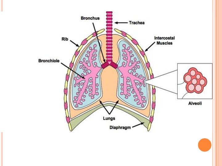
the body in contact with the outside environment. Like
the skin and the gastrointestinal mucosa, the lungs are continuously exposed to a diverse array of microbes and organic and inorganic particulate materials. Innate immune mechanisms defend the air spaces from the array of microbial products that enter the lungs on a daily basis and are evident from the nasopharynx to the alveolar membrane. Particles 1 min size and smaller, the size of bacteria and viral particles, are carried to the alveolar surface where they interact with soluble components in alveolar fluids (e.g., IgG, complement, surfactant, and surfactant-associated proteins) and alveolar macrophages. Normally, alveolar macrophages account for approximately 95% of airspace leukocytes, with 1 to 4% lymphocytes and only about 1% neutrophils, so that the alveolar macrophage is the sentinel phagocytic cell of the innate immune system in the lungs.
The soluble constituents of airway and alveolar fluids have an important role in innate immunity in the lungs. In the conducting airways, constituents of airway aqueous fluids include lysozyme, which is lytic to many bacterial membranes; lactoferrin, which excludes iron from bacterial metabolism; IgA and IgG; and defensins, which are antimicrobial peptides released from leukocytes
and respiratory epithelial cells. IgG is the most abundant immunoglobulin in alveolar fluids, and complement proteins and surfactant-associated proteins serve as additional microbial opsonins.
Слайд 12 Alveolar macrophages are long-lived, with a turnover rate
of only approximately 40% in 1 year. By contrast,
substantial turnover of both lung tissue and peritoneal macrophages occurs within a period of 21 days.Alveolar macrophages are avidly phagocytic and ingest all types of inhaled particulates that reach the alveolar spaces.
Remarkably, one of the primary roles of the alveolar macrophage is to keep the airspaces quiet, and they ingest large numbers of inert particulates like amorphous silicates and carbongraphite particles without triggering inflammatory responses. Normally, the airspace environment is a relatively quiet place despite the array of microbial and other products that enter the airspaces by inhalation or subclinical oropharyngeal aspiration.
Alveolar macrophages are the masters of contradictory function. They are essential for steady-state ‘hoovering’ of daily cellular debris but are also ideally placed to initiate a strong inflammatory response to something more pathogenic. How do alveolar macrophages so rapidly distinguish between these two functions?
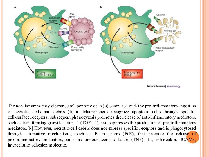
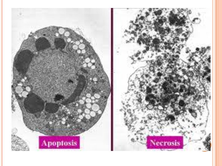
Слайд 13 The non-inflammatory clearance of apoptotic cells (a) compared
with the pro-inflammatory ingestion of necrotic cells and debris
(b). a | Macrophages recognize apoptotic cells through specific cell-surface receptors; subsequent phagocytosis promotes the release of anti-inflammatory mediators, such as transforming growth factor- 1 (TGF- 1), and suppresses the production of pro-inflammatory mediators. b | However, necrotic-cell debris does not express specific receptors and is phagocytosed through alternative mechanisms, such as Fc receptors (FcR), that promote the release of pro-inflammatory mediators, such as tumour-necrosis factor (TNF). IL, interleukin; ICAM3, intercellular adhesion molecule.Слайд 18 In healthy individuals, the airspaces are replete with
mechanisms that prevent an inflammatory response from occurring. This
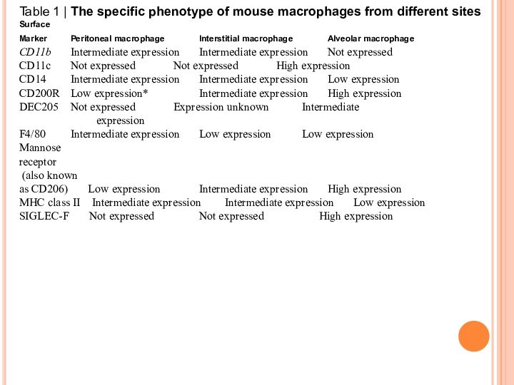
in turn affects the function and the phenotype of the alveolar macrophages, which are one of the few cell populations to reside in the healthy airspaces, in addition to a small number of lymphocytes. Alveolar macrophages from mice have been shown to be poor at presenting antigens to T cells, although they are capable of transporting antigens to the lung-draining lymph nodes. Human alveolar macrophages also induce T cell antigen-specific unresponsiveness as a result of poor antigen presentation and a lack of expression of co‑stimulatory molecules, such as CD86; this promotes tolerance to innocuous antigens. In addition, alveolar macrophages show decreased phagocytic activity compared with lung interstitial macrophages and also have a reduced respiratory burst. Furthermore, they produce immunosuppressive prostaglandins and transforming growth factor‑β (TGFβ), which suppress T cell activation. Alveolar macrophages may drive the development of forkhead box P3 (FOXP3)+ regulatory T cells by secreting TGFβ and retinoic acid, although recent evidence suggests that tissue-resident macrophages in the lungs can also secrete these molecules.Слайд 19 Table 1 | The specific phenotype of mouse
macrophages from different sites
Surface
Marker Peritoneal macrophage Interstitial
macrophage Alveolar macrophage CD11b Intermediate expression Intermediate expression Not expressed
CD11c Not expressed Not expressed High expression
CD14 Intermediate expression Intermediate expression Low expression
CD200R Low expression* Intermediate expression High expression
DEC205 Not expressed Expression unknown Intermediate expression
F4/80 Intermediate expression Low expression Low expression
Mannose
receptor
(also known
as CD206) Low expression Intermediate expression High expression
MHC class II Intermediate expression Intermediate expression Low expression
SIGLEC-F Not expressed Not expressed High expression
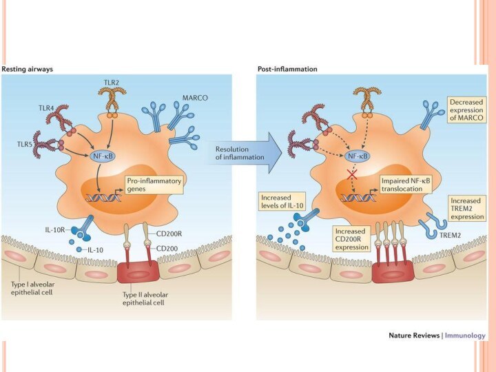
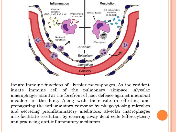
Слайд 20 Innate immune functions of alveolar macrophages. As the
resident innate immune cell of the pulmonary airspace, alveolar
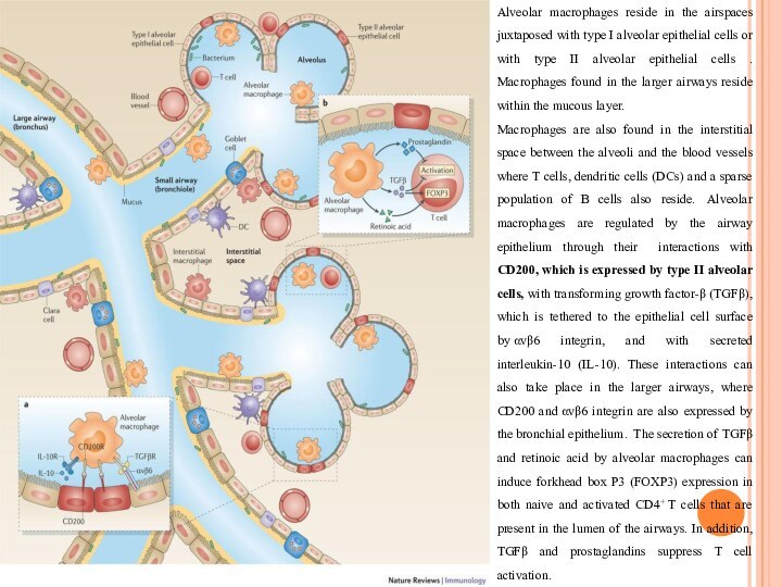
macrophages stand at the forefront of host defence against microbial invaders in the lung. Along with their role in effecting and propagating the inflammatory response by phagocytosing microbes and secreting proinflammatory mediators, alveolar macrophages also facilitate resolution by clearing away dead cells (efferocytosis) and producing anti-inflammatory mediators.Слайд 21 Alveolar macrophages reside in the airspaces juxtaposed with
type I alveolar epithelial cells or with type II
alveolar epithelial cells . Macrophages found in the larger airways reside within the mucous layer.Macrophages are also found in the interstitial space between the alveoli and the blood vessels where T cells, dendritic cells (DCs) and a sparse population of B cells also reside. Alveolar macrophages are regulated by the airway epithelium through their interactions with CD200, which is expressed by type II alveolar cells, with transforming growth factor-β (TGFβ), which is tethered to the epithelial cell surface by αvβ6 integrin, and with secreted interleukin-10 (IL-10). These interactions can also take place in the larger airways, where CD200 and αvβ6 integrin are also expressed by the bronchial epithelium. The secretion of TGFβ and retinoic acid by alveolar macrophages can induce forkhead box P3 (FOXP3) expression in both naive and activated CD4+ T cells that are present in the lumen of the airways. In addition, TGFβ and prostaglandins suppress T cell activation.


